Sample is a small quantity of material taken from an object or body of matter for laboratory examination and analysis. Sampling involves selection of a minor quantity from a large bulk with the purpose of evaluating the lot. The method of collecting or obtaining and handling of samples are very crucial or critical in laboratory and diagnostic investigations.
Specimen is used to refer to materials collected from humans for Laboratory research or practical exercises. For any microbiological examination the proper collection and transportation of an appropriate sample to the laboratory should be of first priority. Sample containers should be sterile and labelled with date, time, location and other necessary information or identity. Samples collected from distant places should be transported in appropriate medium and containers. Samples should be refrigerated at preferably, 0-4°C from time of collection.
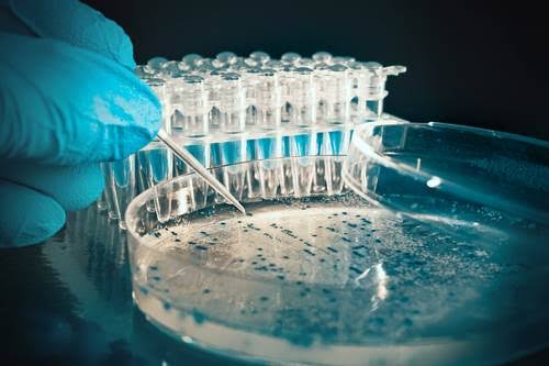
CULTURING OF SPECIMENS
This is the deliberate introduction of a microbiological material or specimen (inoculum) on or into a solid or liquid microbiological medium. The purpose of culturing Specimens is for encouraging the growth and proliferation of microbial cells in the inoculum.
It serves also to demonstrate the presence of organisms which may be causing disease. The technique of culturing is used to test the sensitivity of pathogens to antimicrobial agents. Several methods are used for culturing or inoculating a sample or specimen on the appropriate medium. These methods include Streaking, pour plate, Spread plate, Drop count, most probable number technique.
PREPARATION OF STOCK CULTURES
Before proceeding to identify a microorganism, it iss essential to ensure that enough of the culture is available for use. This is done by the preparation of stock cultures, Various methods are available for stocking Bacteria and include the use of slants. This is the most commonly used method and involves placing the bacterium on agar slants and storing the slants in the refrigerator. Many bacteria will remain viable on such slants for weeks or even months. Other bacteria will require to be transferred onto fresh agar slants at weekly or even daily intervals for them to survive.
UBIQUITY OF MICROORGANISMS
Microorganisms (bacteria, fungi, viruses) exist everywhere in nature. They interact with the non-living environment wherever they are found to form an ecological system. Some are found in almost every environmental niche. They are present in soil, water and air, in most kinds of inorganic or organic non living matter as well as in vast numbers within and on the surface of all living creatures.
They can also be found in the different parts of the human body such as the skin, nose, mouth, intestinal and urinogentl
al tracts. The internal organs and tissues of man are normally sterile (no organisms grow there).
The laboratory is filled with a lot of microorganisms suspended in the air, laboratory benches, and other surfaces. With this knowledge, the laboratory technologist must practice techniques that will not allow these organisms contaminate his or her working materials eg media, equipment etc.
DEMONSTRATION OF UBIQUITY OF MICROORGANISMS
Materials:
10 -15 nutrient agar plates, SDA, grease pencil, sterile swab sticks.
Procedure
1.Uncover two sets of plates (4) and expose them on the laboratory bench surface and floor of the laboratory for about 10 and 30 minutes respectively. Cover the plates and label them A and B respectively.
2. Uncover another two sets of plates and expose them on a stool for 10 and 30 minutes respectively. At the end of the time cover the plates and label them C and D respectively.
3. Collect little soil from outside the laboratory and inoculate onto 2 plates Cover the plates and label them E.
4. Gently rest your left and right fingers on 2 agar plates, cover the plates and label F.
5. Let the sleeve of your lab coat touch the agar surfaces. Cover the plates and label G.
6. Using sterile swab sticks, swab the skin, nose, and ears of the laboratory staff and some students. Inoculate onto nutrient agar plates in duplicate and label H, I and J respectively.
7. Incubate all the plates at 37°C for 24 h, record the growth of organisms on the plates, count the colonies,
EXAMINATION OF UBIQUITY PLATES
The macroscopic and microscopic identification of the Various colonies observed will serve as preliminary criteria for identification of the organisms into appropriate genera and species.
The macroscopic identification of the organisms reveals the shape, size, texture, colour and pigmentation of the organism. While the gram reaction reveals the cellular arrangement, shape of bacterium and other characteristics such as spore and capsule formation.
Microbiological Sampling Of Air
There are various methods of microbiological air sampling namely settle plates, slit sample and cascade impaction sampler.
Settle Plates:
In the past, the procedure frequently adopted for determining the relative numbers and species of microorganisms present in air has been to expose or open plates of culture medium for given periods of time e.g ½ or 1h.
A count of the colonies after incubation of the plates for 14h at 37°C yields a relative estimate of the number of organisms present. And if blood agar is used, the occurrence in the air of pathogenic staphylococci and streptococci can be determined.
This method has proved valuable in demonstrating the presence of such organisms in air dust.
Slit Sampler:
It is recognized that the simpler method of exposing plates has certain limitations as a means of studying the bacteriology of air. For example, it is not a satisfactory method of detecting bacteria in very small suspended particles such as droplet-nuclei.
More elaborate procedures have therefore been adopted. A technique introduced by Bourdillon et al (1914) involves the use of a social instrument the “slit sampler” by which known volume of aur is directed onto a plate through a slit 0.25mm wide, the plate being mechanically rotated ea that the organisms are evenly distributed over it.
One cubs foot of air per minute is allowed to pase through the slit and samples of 1 to 10 cubic feet or more may be tested More advanced models of slit sample have a timing arrangement that allows the number of colonies on each sector of the plate to be related to the number of bacteria – carrying particles sampled in a particular part of the sampling period.
CASCADE IMPACTION SAMPLER
The cascade impaction sampler consists of four impaction stages and an after-filter that. allows the separation and collection of airborne particles in five size ranges. These ranges include 257 um, 1.0 um, 0.5 um and 0.25 um and 2.0 um after-filter. Size fractionated samples can be analyzed gravimetrically, chemically and microbiologically.
LEVELS OF AIR INFECTION
These are generally expressed in terms of the count of bacterial colonies of all kinds made on blood or nutrient agar plates incubated for 24h at 37°C. When plates are incubated for a much longer period at room temperature, the counts are often very much higher as a result of the slow growth of saprophytic organisms that do not grow well at 37°C.
The counts are expressed as the number of bacteria carrying – particles per cubic foot of air when the examination is made with a slit sampler and as the number of bacteria carrying particles settling on a petri dish per minute per hour when it is made with settle plates.
Under conditions of normal occupation, the air in hospital wards, offices, schools and private houses commonly shows levels of contamination in the range of 5 to 100 bacteria carrying particles per 3” in settle plates per minute.The great majority of the bacteria found in the air are harmless saprophytes or commensals.